Get the latest news delivered monthly to your inbox. Subscribe to The Cancer Code.
Subscribe NowCancer Discoveries
The latest cancer discoveries and innovations coming out of cancer research labs at the Medical College of Wisconsin.

The Ojesina Lab Targets Cervical Cancers Prevention Can't Catch
By studying how genetic changes interact with viruses and bacteria inside tumors, researchers are working to uncover what sets these cancers apart and how that knowledge could lead to better care.

From Molecules to Medicine: How Data Powers Precision Oncology
A connected cycle of discovery, validation, prediction, and personalization is transforming molecular insights into more precise and effective cancer therapies.

MCW Takes Cancer Care by Storm: Theranostics and the 'Next Wave' of Precision Oncology
By using specialized imaging to reveal where cancer is active, doctors can target therapy directly to tumors in the same step, transforming the way cancer is detected, tracked, and treated.

Global Study Tests Next-Generation HER2 Therapy for Metastatic Breast Cancer
A promising phase 3 trial is testing a next-generation therapy designed to help patients whose cancer has stopped responding to standard treatments, with the goal of offering better control of the disease and improving quality of life.

A ‘Personalized Pharmacy’: Cell Therapy Shared Resource Engineers Lifesaving Treatments for Cancer and Beyond
Researchers at MCW are pushing cell therapy into new frontiers, exploring its potential in neurological disorders, genetic diseases, and other serious conditions.

Drugging the Undruggable: McFall Lab Unlocks New Treatments for KRAS-Driven Cancers
By challenging long-held assumptions about how certain KRAS mutations function, the McFall Lab is uncovering new ways to treat some of the most aggressive and difficult-to-treat cancers.

MCW Cancer Center Trial Tests Cutting-Edge Radioimmunotherapy to Improve AML Outcomes
Researchers at MCW are testing a more precise treatment approach that delivers radiation directly to cancer cells while leaving healthy tissue unharmed.

Researchers Use Genetic Testing to Identify More Effective Treatments for Advanced Prostate Cancer
Scientists found that platinum-based chemotherapy may help men with advanced prostate cancer who no longer respond to PARP inhibitors, especially those with BRCA2 mutations.

MCW Co-Designed Trial Offers Safer Treatment to Vulnerable Patients with Head and Neck Cancer
Results of the NRG-HN004 study help to establish a new standard of care while addressing a critical gap in cancer treatment.

MCW Cancer Investigators Play Key Role in FDA Approval of First T-Cell Therapy for Solid Tumors
Approved for adults with aggressive synovial sarcoma, afami-cel provides a novel treatment approach for this challenging cancer. The Center plans to be the first in Wisconsin to offer it to patients.

A Catalyst for Discovery: MCW’s World-Class Brain Bank Drives Groundbreaking Neuro-Oncology Research
The Brain Bank is rapidly evolving into a hub for neuro-oncology-focused research that equips MCW scientists with unique equipment and an expansive database to accelerate breakthroughs and bring hope to patients with brain cancer.

MCW Scientists Unveil a New More Tolerable Treatment Option for Patients with Thyroid Cancer
Investigators discovered a novel combination of two drugs was powerful in killing thyroid cancer cells with RET mutations and resulted in fewer adverse effects than existing treatments.

MCW Researchers Use Cutting-Edge Technology to Enhance Immunotherapy in Head and Neck Cancers
A groundbreaking study showed that special T cells capable of recognizing and killing cancer cells could be taken out of patients’ tumors and made stronger to fight their disease.

MCW Scientists Identify Patients with Pancreatic Cancer Who May Respond to Immunotherapy
A new study has identified four distinct subtypes of pancreatic cancer, revealing that some types have a better outlook and the potential to respond better to immunotherapy.

Mito-ATO Makes Immunotherapy More Potent Against Tumors, 'Shows Great Clinical Potential'
Collaborative research shows the MCW-created drug, Mito-ATO, may play a crucial role in improving cancer treatment by altering and reprogramming immune cells.

MCW Cancer Scientists Make a Breakthrough in Treating Aggressive Uterine Cancer
A new study is the first ever to show the anti-cancer therapeutic potential of using drugs selinexor and eribulin in uterine leiomyosarcoma.

MR Tumor Regression Grade Can Help Oncologists Guide Treatment for Patients with Rectal Cancer
A new study is showing that when used alone, magnetic resonance (MR)-based imaging for grading tumors in patients with rectal adenocarcinoma is an insufficient tool for measuring pathologic complete response (pCR).

Scientists Identify FOXM1 Protein as a Key Driver of Myeloma Metabolism
In a recent study, investigators take a closer look at the metabolic role of genes to determine the genetic and biological pathways associated with high-risk myeloma and relapsed/refractory myeloma. The research opens the door to new therapies and prevention.

Scientists Design Integrin Inhibitor Principle to Aid the Development of Drugs Targeting Cancer
Integrins are molecules that play an important role in cancer cell migration and survival. In a recent study, investigators developed a new design for improving the safety and efficacy of cancer treatments as well as treatments for autoimmune disease and thrombosis.
Clinical Trials
Featured studies paving the way for new treatments and lifestyle interventions for patients with cancer.

Cilta-Talq Fusion Study Examines a New Bridging Approach to CAR T-Cell Therapy in Multiple Myeloma
Dr. Othman Akhtar uses targeted immunotherapy to stabilize patients while their personalized CAR T-cell therapy is manufactured, supporting safer and more effective treatment.

Early-Phase Trial Explores a New Immunotherapy Approach for Hard-to-Treat Solid Tumors
A first-in-human study is testing a targeted IL-2-based therapy designed to boost the body’s own cancer-fighting cells.

MCW Cancer Center Expands Access to Precision Oncology and Rare Cancer Care
Under the leadership of Dr. Razelle Kurzrock, more patients than ever will benefit from personalized, next-generation cancer treatments.

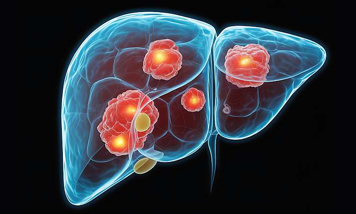
ROUTE 90 Trial Brings Cutting-Edge Radiation Therapy to Patients with Liver Cancer
A multi-center trial is testing a next-generation therapy that delivers targeted radiation directly to tumors with greater precision and visibility than ever before.

New Study Examines Long-Term Impact of Next-Generation BTK Inhibitor in Blood Cancers
MCW Cancer Center clinical research teams activated a phase 4 study to explore the long-term use of pirtobrutinib, a therapy for chronic lymphocytic leukemia.

Beyond the Beam How MCW Innovators Are Transforming Radiation Therapy With Sonoptima
The first-of-its-kind wearable device uses AI and ultrasound to monitor bladder fullness in real time, helping patients with pelvic cancers know exactly when to start radiation treatment.

The Science of Survivorship Every Day Counts Study Improves Life for Women with Metastatic Breast Cancer
The 16-week lifestyle intervention aims to understand what happens when women with breast cancer make small, manageable changes to how they eat, move, and manage their health.

MASTER-2 Trial Advances Personalized Treatment Approach for Multiple Myeloma
Findings from the study could guide care decisions and reduce unnecessary treatment for patients who are already doing well.

Turning Science into Survivorship: Children’s Wisconsin Researchers Drive Breakthroughs in Pediatric Cancer Care
The MACC Fund has turned community generosity into life-changing childhood cancer research at MCW and Children’s Wisconsin.

MCW Cancer Investigators Evaluate New Targeted Therapy for Advanced Leiomyosarcoma
The global trial aims to determine if this novel approach helps control tumors longer than standard treatment alone.

MCW-led Trial Tests Novel Combination Treatment for Myelodysplastic Syndrome
The phase 1 trial tests the safety and best dose of iadademstat with azacytidine, aiming to improve treatment response and slow disease progression.

New Children's Wisconsin Trial Tests Immunotherapy with Chemotherapy in High-Risk Neuroblastoma
This novel approach may target cancer cells more effectively, potentially reducing relapse and improving survival for patients experiencing one of the most aggressive pediatric cancers.

MCW Cancer Center Launches MyeloMATCH to Advance Precision Medicine for Myeloid Cancers
The phase 2 study identifies genetic mutations driving aggressive myeloid cancers, enabling doctors to provide more precise and effective therapies tailored to each patient.

MCW Cancer Center Welcomes New Leadership to Improve Access to Cutting-edge Clinical Trials
Building on its commitment to improve health for all, the Center has appointed Dr. Callisia Clarke as Co-Associate Director of Clinical Research with a focus on clinical trial equity.

Trailblazers in Innovation: MCW Researchers Harness the Power of AI to Improve Clinical Trial Matching
A new pilot study conducted by researchers at the MCW Cancer Center and CTSI automates patient-trial matching by using a large language model (LLM), a Triomics platform called OncoLLM, to analyze vast amounts of data in a fraction of the time it would take a human, and with greater precision.
Learn how we’re setting new standards for clinical trial equity

MCW's CAR T-Cell Lab Helps Scientists Bring Lifesaving Therapies to Patients with Blood Cancers
Cancer investigators recently treated the 100th patient in an ongoing clinical trial that uses an in-house manufactured, dual-targeted CAR-T therapy for patients with B-cell malignancies.

Center Leads Groundbreaking Change in Medicare Policy for MDS Patients in Need of Transplants
Decades-long research from MCW Cancer Center investigators has led to a Medicare policy change that expands coverage for certain stem cell transplants for patients with myelodysplastic syndromes.

Patient Navigation Program Improves Access to Life-Saving Clinical Trials
In a significant stride towards inclusivity in cancer care, the MCW Cancer Center has launched a new program to improve clinical trial access in underserved communities.

PROTECT-PANC Trial Evaluates Precision Medicine Strategies in Patients with Pancreatic Cancer
The fast-track trial bolsters MCW’s dedication to advancing precision medicine strategies across a wide spectrum of cancer types.

JANUS Trial Improves on Past Treatment Strategies for Advanced Stage Rectal Cancer
Investigators say the phase 2 study is “transformative” because it allows patients to be managed with a watch and wait approach.

Translational Research Unit Celebrates 10 Years of Accelerating Novel Cancer Therapies
The state-of-the-art clinical and research facility has been instrumental in advancing innovative ideas and launching practice-changing cancer clinical trials.

Novel Treatment Strategy Offers New Hope for Young Patients with Classic Hodgkin Lymphoma
In a recent trial, MCW investigators tested a new salvage therapy approach that incorporated targeted therapy, immunotherapy, and chemotherapy that may be safer and more effective than the standard of care for treating pediatric patients with relapsed cHL.

Combination of Chemotherapy and CAR-T Therapy Helps Leukemia Patients Achieve Remission
MCW Cancer Center investigators discovered that chemotherapy coupled with CAR-T infusion helped patients with acute lymphocytic leukemia achieve another remission, opening the door to promising new therapies.

CAR-T Therapy Slows Multiple Myeloma in Patients Who Have Stopped Responding to Other Treatments
MCW’s Binod Dhakal, MD, presented the first results from the Phase 3 CARTITUDE-4 trial, which showed that cilta-cel reduced the risk of MM progression or death by approximately 74%.

Patients Receive "Rockstar" Treatment for Chronic Graft-Versus-Host Disease
MCW Cancer Center scientists conducted a pivotal trial leading to the expedited approval of REZUROCK, a ground-breaking new therapy available to adult and pediatric patients that works by rebalancing a patient’s immune system.
Cancer in Our Community
Research insights and policies aimed at improving the life and well-being of people across Wisconsin and beyond.

The Health Griots Program Has Turned Prostate Cancer Awareness into Action
What started as a small group of prostate cancer survivors sharing their stories has grown into a brotherhood, and a movement, that is changing how Black men across Milwaukee talk about prostate cancer.

Immersive Cancer Center-UWM Training Program Gives Future Scientists a Head Start in Discovery
Through the UW-Milwaukee Undergraduate Research Experience Program, what begins as curiosity becomes confidence as students step into MCW Cancer Center labs, where mentors and hands-on research experiences open doors and set them on a path toward impactful careers in cancer science.

Inaugural Audaxity Ride Raises $1 Million to Accelerate Cancer Research at the MCW Cancer Center
Funds from the community ride will help turn discoveries into new cancer treatments, train future physician-scientists, and expand community-driven research efforts.

Translating Hope: New Mural Connects Science and Community at the Center for Cancer Discovery
Artist Mauricio Ramirez’s mural transforms the building’s main conference space into a vibrant symbol of unity, depicting how science and community come together in the fight against cancer.

Cancer from Every Angle: GEO Shared Resource Helps Researchers Turn Data into Action
The Geospatial, Epidemiology, and Outcomes Shared Resource helps researchers identify community patterns with patient-level insights to advance cancer research and care.

The Breakthrough Ride: More Than 1,000 Participants Join Inaugural Audaxity Event to Fuel Cancer Discoveries
With the support of founding sponsor Associated Bank, 100 percent of the funds raised through Audaxity will fuel new cancer discoveries.

Support That Changes the Story: How Patient Navigation Expands Access to Lifesaving Clinical Trials
Here, patients facing the uncertainty of a cancer diagnosis don’t have to navigate the journey alone. Patient navigation helps people across our communities gain access to clinical trials that could improve their outcomes.

MCW Cancer Center Hosts NCI Site Visit, Marking a Major Milestone on the Road to Designation
This moment brings the Center closer to unlocking the potential for increased federal funding, expanded research collaborations, and faster translation of scientific discoveries into care for the 3.4 million people it serves.

Audaxity Bike Giveaway Helps More Riders Join the Movement to End Cancer
Wisconsin cancer survivors and caregivers were gifted 25 new bicycles from Associated Bank in advance of the inaugural Audaxity Bike Ride.

Lubna Chaudhary Awarded Pilot Funding to Unlock the Secrets of Endocrine Resistance in Breast Cancer
The Our Patient Project study lays the foundation for more personalized treatment strategies, ensuring patients can receive therapies suited to their cancer’s unique biology.

Cancer Prevention Starts With Grassroots Efforts Led by the Community Outreach and Engagement Team
The small but mighty team delivers a wide range of services—from health education to screenings to patient navigation—directly into the community, especially in underserved neighborhoods.

Wisconsin's First Compact Proton Therapy System: Froedtert & MCW Forging a New Path in Cancer Care
In summer 2025, the Froedtert & the Medical College of Wisconsin health network will begin offering a game-changing form of radiotherapy for patients in our community and across the state.

MCW Blood Cancer Researchers Lead Efforts to Reduce Disparities in Access to Lifesaving Treatment
A new study reveals barriers underserved patients face in accessing stem cell transplants, laying a foundation for developing targeted programs that advance health equity.

Health Griots Program Empowers Prostate Cancer Survivors to Lead the Charge in Eliminating Disparities
By educating on cancer prevention, early detection, and access to care, the Health Griots are working to close gaps in support and connect underserved patients with the care they need to live long, healthy lives.

United Against Cancer MCW Researchers Join Forces with the Community to Eliminate Disparities
The Research & Community Scholars Program creates a space for fair and trusting relationships to develop between MCW and the community, helping to ensure that MCW Cancer Center research is both effective and equitable.

MCW Scientists Discover Chemokine Levels May Contribute to Prostate Cancer Disparities
A new study reveals that biological factors may explain racial disparities in prostate cancer lethality; the findings could help guide therapeutic approaches to improve outcomes in African American men.

Transgender and Nonbinary People, and Providers, Benefit from Better Awareness of Screening Guidelines
Screenings help catch cancer early, resulting in more promising health outcomes. While the transgender and nonbinary population is growing, the majority are unsure of when and whether to be screened. A new study points to the need for greater cancer education and care.

Dr. Elizabeth Hopp Awarded Pilot Funding to Improve Outcomes for Patients with Rare Ovarian Cancer
The MCW Cancer Center has awarded Dr. Elizabeth Hopp an Our Patient Project pilot grant for her innovative research that may drive discovery of more effective treatments for ovarian cancer.

MCW’s Commitment to Advancing Precision Medicine ‘Levels the Playing Field’ for Patients with Cancer
A panel of MCW Cancer Center leaders share how the institution is harnessing cutting-edge precision medicine techniques at the 11th annual ACS CAN Cancer Research & Innovation Forum.
See how we bring the right treatments to the right patient at the right time
People and Progress
Recent leadership announcements, program updates, and Cancer Center milestones.

Precision Under Pressure: Dr. David Seo Helps Guide Urgent Treatment Decisions in Pancreatic Cancer
For patients with pancreatic cancer, timing is everything. Dr. David Seo harnesses precision medicine to match therapies to each tumor, faster and smarter.

Jennifer Knight Appointed Co-Leader of Cancer Control Program
A leading voice in psycho-oncology, Dr. Knight investigates how factors like stress impact the body—with the goal of improving emotional and physical well-being for patients.

Breakthroughs Beyond the Bench: Miller Lab Turns Breast Cancer Discoveries into Better Care
Todd Miller, PhD, Wisconsin Breast Cancer Showhouse Endowed Professor of Breast Cancer Research, is dedicated to unraveling one of cancer’s most complex puzzles: why breast cancer adapts, resists treatment, and returns long after therapy ends.

From the Gut to the Genome: Kudek Lab Engineers Better Treatments for Children with Cancer
Guided by science and collaboration, Dr. Matthew Kudek is driven to transform complex immune responses into solutions that protect children long after cancer is gone.

Cancer Ends Here: MCW Celebrates the Grand Opening of the Center for Cancer Discovery
The new cancer research facility will stand as a hub where bold ideas, collaborative partnerships, and community voices unite to transform cancer prevention, diagnosis, treatment, and survivorship for generations to come.

Advancing a Healthier Wisconsin Supports Cancer Research and Improves the Health of Our Community
AHW-supported research plays a vital role in solving the most pressing public health challenges and shaping a healthier future for families across the state.

Celebrating Clinical Trials Day: Voices from the Frontlines of Cancer Research
Clinical research professionals are at the heart of medical progress, helping to guide patients through complex care journeys.

A New Era: MCW Center for Cancer Discovery
From breaking ground to breaking through, the MCW Center for Cancer Discovery stands as a beacon to scientific discovery.

Faculty Feature: Vanessa Leone, PhD
Leveraging computational approaches such as artificial intelligence, Dr. Leone’s research aims to uncover new and innovative cancer treatments.

Marilyn Larson, MBA: Shaping the Future of Cancer Care
A Wisconsin native, Marilyn joined MCW in 2014, driven by a mission to further establish the Cancer Center as a leader in accessible, innovative cancer research and care.

Mary Horowitz, MD, MS: An Unexpected Path that Changed Cancer Care
Dr. Horowitz is a world-renowned leader in blood and marrow transplantation research that has helped improve survival rates and made transplants safer and more widely accessible.

Razelle Kurzrock, MD, FACP: Redefining What’s Possible
Dr. Kurzrock is helping to transform the landscape of cancer research and care to achieve more precise diagnoses, personalized and targeted therapies, and improved patient outcomes.

Faculty Feature: Fumou Sun, PhD
Dr. Sun is dedicated to translating CAR-T cell therapies into clinical trials, bridging the gap between innovative laboratory research and transformative patient care.

Audaxity Bike Fundraiser Welcomes New Leadership to Advance Goals, Raise Millions for Cancer Research
Lauren “LB” Bennett’s arrival marks an exciting new chapter for Audaxity as she pioneers the development of the inaugural ride, and brings together a community boldly united to go after the Center’s vision of eradicating cancer.

2nd Annual Trainee Symposium Celebrates Learners Pushing Past Barriers to Unlock Cancer Discoveries
More than 170 attendees gathered at the Milwaukee County Zoo’s Zoofari Conference Center to recognize and learn from next-generation cancer scientists.

Cancer Research Forum Celebrates Its First Year of Driving Scientific Collaboration and Innovation
The collaborative meeting and social event creates a space for Cancer Center members to share unpublished research and gather quality feedback from peers.

Faculty Feature: Todd Miller, PhD
Dr. Miller’s lab aims to understand the causes of drug resistance in breast cancer, and to develop improved treatment strategies to prevent and overcome it.

Faculty Feature: Staci Young, PhD
In her new role as associate director of community outreach and engagement, Dr. Staci Young will lead the team responsible for advancing clinical, research, and policy initiatives that extend the center’s reach with under-resourced groups, and improve access to cancer prevention, life-saving clinical trials, and survivor support groups.

Partnership with UW-Milwaukee Aims to Foster a Lifetime Commitment to Science
The MCW Cancer Center recently awarded 10 Wisconsin students $100,000 in scholarships to pursue cancer research careers. The educational program provides an immersive experience that enables students to work alongside nationally-recognized scientists.
